Ct Anatomy Pelvis Muscles : How To Read A Ct Of The Abdomen And Pelvis Radiology Case Radiopaedia Org / There are many muscles that form the pelvic floor, including puborectalis, pubococcygeus, iliococcygeus and coccygeus.
byAdmin•
0
Ct Anatomy Pelvis Muscles : How To Read A Ct Of The Abdomen And Pelvis Radiology Case Radiopaedia Org / There are many muscles that form the pelvic floor, including puborectalis, pubococcygeus, iliococcygeus and coccygeus.. It is the most complete reference of human anatomy available on web, ipad, iphone and android devices. Axial section through male bladder. There are many muscles that form the pelvic floor, including puborectalis, pubococcygeus, iliococcygeus and coccygeus. The muscle originates from the body of the pubis and attaches to the pectineal line and proximal part of the linea aspera of femur. Ct mri radiographs anatomic diagrams and nuclear images.
There are many muscles that form the pelvic floor, including puborectalis, pubococcygeus, iliococcygeus and coccygeus. Radiographers suggest an abdominal ct scan to look for the following: Ap pelvis anatomy radiographs ct protocols pelvic ring fx ap force lateral vertical acetabular fx walls columns letournel judet view wow Int/ext obliques transversus spine muscles: The thigh has some of the body's largest muscles.
Cross Sectional Anatomy Of The Male Pelvis Springerlink from media.springernature.com Male abdomen and pelvis ct scan form no 7. Suspensory ligament of the penis. The ct appearance of the normal ligamentous, vascular, and visceral anatomy of the female pelvis can be confusing. Int/ext obliques transversus spine muscles: They form a large sheet of skeletal muscle that is thicker in some areas than in others. Thigh muscles are responsible for allowing normal gait and proper lower extremity function (1). Anatomy ct axial abdomen and pelvis male from 3.bp.blogspot.com the lateral superficial muscles, the transversus and external and internal oblique muscles, originate on the rib cage and on the pelvis (iliac crest and inguinal ligament) and are attached to the anterior and posterior layers of the sheath of the rectus. The muscles of the pelvic floor are collectively referred to as the levator ani and coccygeus muscles.
Anatomy ct axial abdomen and pelvis male from 3.bp.blogspot.com the lateral superficial muscles, the transversus and external and internal oblique muscles, originate on the rib cage and on the pelvis (iliac crest and inguinal ligament) and are attached to the anterior and posterior layers of the sheath of the rectus.
Thigh muscles are responsible for allowing normal gait and proper lower extremity function (1). Explore over 6700 anatomic structures and more than 670 000 translated medical labels. Abdominal computed tomography (ct) is a type of medical imaging procedure used to diagnose and monitor internal stomach issues, like cancer, bowel obstruction, and abdominal pain. Muscles that attach from the pelvis to the trunk and cross the lumbosacral joint muscles that attach from the pelvis to the thigh/leg and cross the hip joint pelvic floor muscles that are located wholly within the pelvis The muscles of the pelvic floor are collectively referred to as the levator ani and coccygeus muscles. These two muscles are primarily responsible for maintaining pelvic stability when one foot is lifted off the ground (e.g., during the swing phase of walking). The muscle originates from the body of the pubis and attaches to the pectineal line and proximal part of the linea aspera of femur. This mri male pelvis axial cross sectional anatomy tool is absolutely free to use. It is a basin shaped muscular diaphragm that helps to support the visceral contents. The bony pelvis is formed by the sacrum and coccyx and a pair of hip bones ossa coxae which are part of the appendicular skeleton. Use the mouse scroll wheel to move the images up and down alternatively use the tiny arrows (>>) on both side of the image to move the images.>>) on both side of the image to move the images. Explore anatomie's range of luxury travel clothes for women at anatomie store. Pelvic muscles ct anatomy and ct scan of the abdomen and pelvis shows a normal appendix 7 pelvic muscles ct anatomy pelvic muscles ct anatomy and ct scan of the abdomen and pelvis shows a normal appendix gallery at human diagram chart.
The medial thigh muscles are responsible for the adduction (movement of a body part toward the body's midline) of the leg. They form a large sheet of skeletal muscle that is thicker in some areas than in others. The muscle originates from the body of the pubis and attaches to the pectineal line and proximal part of the linea aspera of femur. Ct, mri, radiographs, anatomic diagrams and nuclear images. It is the most complete reference of human anatomy available on web, ipad, iphone and android devices.
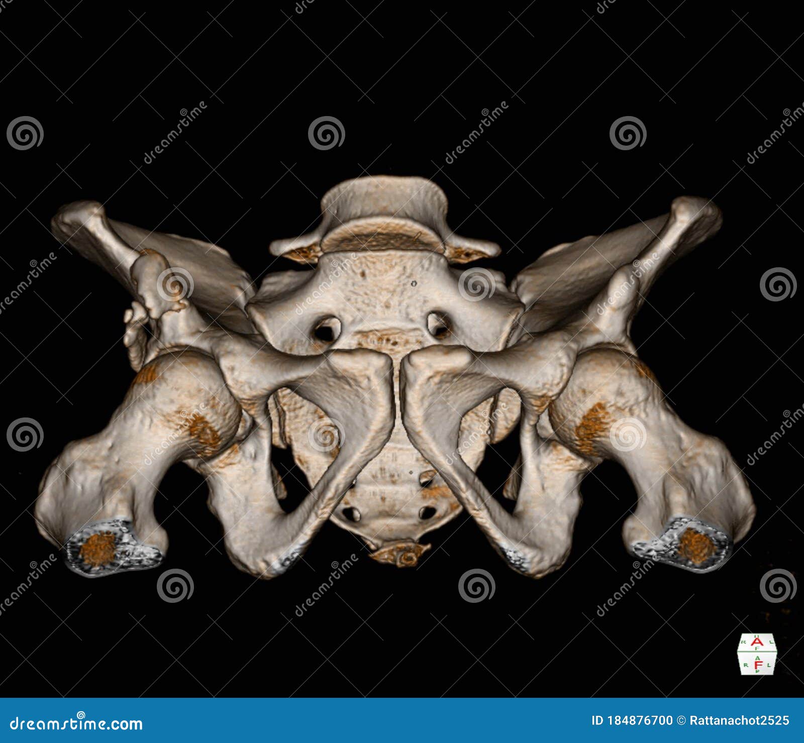
Ct Scan Pelvis Bone 3d Rendering Image Isolated On Black Background Showing There Is A 8mm Thick And 5cm Long Tubular Ossification Stock Illustration Illustration Of Exam Anatomy 184876700 from thumbs.dreamstime.com Pelvic muscles ct anatomy and ct scan of the abdomen and pelvis shows a normal appendix 7 pelvic muscles ct anatomy pelvic muscles ct anatomy and ct scan of the abdomen and pelvis shows a normal appendix gallery at human diagram chart. Ap pelvis anatomy radiographs ct protocols pelvic ring fx ap force lateral vertical acetabular fx walls columns letournel judet view wow Explore anatomie's range of luxury travel clothes for women at anatomie store. Anatomy ct axial abdomen and pelvis male male abdomen and pelvis ct scan form no 1. Functional anatomy of the male pelvic floor online course: Radiographers suggest an abdominal ct scan to look for the following: The full bladder displaces small bowel loops superiorly. There are many muscles that form the pelvic floor, including puborectalis, pubococcygeus, iliococcygeus and coccygeus.
Radiographers suggest an abdominal ct scan to look for the following:
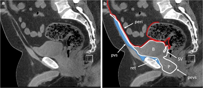
Mr imaging is an excellent tool for noninvasive evaluation of the pelvic floor. It is the most complete reference of human anatomy available on web, ipad, iphone and android devices. Corpus cavernosum of the penis. Ap pelvis anatomy radiographs ct protocols pelvic ring fx ap force lateral vertical acetabular fx walls columns letournel judet view wow The full bladder displaces small bowel loops superiorly. Ct mri radiographs anatomic diagrams and nuclear images. The ct appearance of the normal ligamentous, vascular, and visceral anatomy of the female pelvis can be confusing. The muscles of the pelvis form its floor. This mri male pelvis axial cross sectional anatomy tool is absolutely free to use. An important group of muscles in the pelvis is the pelvic floor. The labeled structures are (excluding the correct side): Computed tomography (ct) remains a valuable technique in the assessment of the female pelvis. Radiographers suggest an abdominal ct scan to look for the following:
The full bladder displaces small bowel loops superiorly. Anatomy ct axial abdomen and pelvis male from 3.bp.blogspot.com the lateral superficial muscles, the transversus and external and internal oblique muscles, originate on the rib cage and on the pelvis (iliac crest and inguinal ligament) and are attached to the anterior and posterior layers of the sheath of the rectus. It is a basin shaped muscular diaphragm that helps to support the visceral contents. Ct mri radiographs anatomic diagrams and nuclear images. Radiographers suggest an abdominal ct scan to look for the following:
Mri Female Pelvis Anatomy Axial Image 22 Pelvis Anatomy Pelvis Anatomy from i.pinimg.com Computed tomography (ct) remains a valuable technique in the assessment of the female pelvis. These two muscles are primarily responsible for maintaining pelvic stability when one foot is lifted off the ground (e.g., during the swing phase of walking). The bony pelvis is formed by the sacrum and coccyx and a pair of hip bones ossa coxae which are part of the appendicular skeleton. The muscles of the pelvis form its floor. There are many muscles that form the pelvic floor, including puborectalis, pubococcygeus, iliococcygeus and coccygeus. It is the most complete reference of human anatomy available on web, ipad, iphone and android devices. Radiographers suggest an abdominal ct scan to look for the following: Use the mouse scroll wheel to move the images up and down alternatively use the tiny arrows (>>) on both side of the image to move the images.>>) on both side of the image to move the images.
Volume rendered display, ct of the pelvis • the external surface of the iliac blade is the attachment site for the gluteus medius and minimus muscles.
Anatomy ct axial abdomen and pelvis male male abdomen and pelvis ct scan form no 1. The full bladder displaces small bowel loops superiorly. Explore anatomie's range of luxury travel clothes for women at anatomie store. Case contributed by assoc prof craig hacking. Thigh muscles are responsible for allowing normal gait and proper lower extremity function (1). Suspensory ligament of the penis. Corpus cavernosum of the penis. Pelvic floor anatomy is complex and is being unraveled by means of magnetic resonance mr imaging. An important group of muscles in the pelvis is the pelvic floor. This mri male pelvis axial cross sectional anatomy tool is absolutely free to use. Male abdomen and pelvis ct scan form no 7. Int/ext obliques transversus spine muscles: The ct appearance of the normal ligamentous, vascular, and visceral anatomy of the female pelvis can be confusing.